| General information about the entry |
| View entry in simple text format |
| Entry name | VIME_MOUSE |
| Primary accession number | P20152 |
| integrated into SWISS-2DPAGE on | September 1, 1998 (release 7) |
| 2D Annotations were last modified on | October 1, 2001 (version 4) |
| General Annotations were last modified on | May 19, 2011 (version 8) |
| Name and origin of the protein |
| Description | RecName: Full=Vimentin;. |
| Gene name | Name=Vim
|
| Annotated species | Mus musculus (Mouse) [TaxID: 10090] |
| Taxonomy | Eukaryota; Metazoa; Chordata; Craniata; Vertebrata; Euteleostomi; Mammalia; Eutheria; Euarchontoglires; Glires; Rodentia; Sciurognathi; Muroidea; Muridae; Murinae; Mus; Mus. |
| References |
| [1] |
MAPPING ON GEL
MEDLINE=96154592; PubMed=8586073; [NCBI, Expasy, EBI, Israel, Japan]
Anderson N.L., Esquer-Blasco R., Hoffmann J.-P., Meheus L., Raymackers J., Steiner S., Witzmann F., Anderson N.G.
''''''An updated two-dimensional gel database of rat liver proteins useful in gene regulation and drug effect studies'';'';''
Electrophoresis 16(1):1977-1981(1995)
|
| [2] |
MAPPING ON GEL
PubMed=11680894; [NCBI, Expasy, EBI, Israel, Japan]
Sanchez J.-C., Chiappe D., Converset V., Hoogland C., Binz P.-A., Paesano S., Appel R.D., Wang S., Sennitt M., Nolan A., Cawthorne M.A., Hochstrasser D.F.
''''''The mouse SWISS-2DPAGE database: a tool for proteomics study of diabetes and obesity'';'';''
Proteomics 1(1):136-163(2001)
|
|
| 2D PAGE maps for identified proteins
|
|
How to interpret a protein
|
BAT_MOUSE {Brown adipose tissue}
Mus musculus (Mouse)
Tissue: Brown adipose tissue
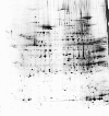
map experimental info
protein estimated location
|
|
BAT_MOUSE
MAP LOCATIONS:
MAPPING (identification):
Peptide mass fingerprinting [2].
|
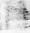
ISLETS_MOUSE {Pancreatic islet cells}
Mus musculus (Mouse)
Tissue: Pancreatic islet

map experimental info
protein estimated location
|
|
ISLETS_MOUSE
MAP LOCATIONS:
MAPPING (identification):
Peptide mass fingerprinting [2].
|
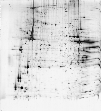
LIVER_MOUSE {Liver}
Mus musculus (Mouse)
Tissue: Liver
map experimental info
protein estimated location
|
|
LIVER_MOUSE
MAP LOCATIONS:
MAPPING (identification):
|
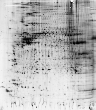
MUSCLE_MOUSE {Gastrocnemius muscle}
Mus musculus (Mouse)
Tissue: Gastrocnemius
map experimental info
protein estimated location
|
|
MUSCLE_MOUSE
MAP LOCATIONS:
MAPPING (identification):
MATCHING WITH THE MOUSE LIVER MASTER GEL [2].
|
NUCLEI_LIVER_MOUSE {Soluble nuclear proteins and matrix from liver tissue}
Mus musculus (Mouse)
Tissue: Liver
map experimental info
protein estimated location
|
|
NUCLEI_LIVER_MOUSE
MAP LOCATIONS:
MAPPING (identification):
Peptide mass fingerprinting [2].
|
WAT_MOUSE {White adipose tissue}
Mus musculus (Mouse)
Tissue: White adipose tissue
map experimental info
protein estimated location
|
|
WAT_MOUSE
MAP LOCATIONS:
MAPPING (identification):
|
| Copyright |
| This SWISS-2DPAGE entry is copyright the Swiss Institute of Bioinformatics. There are no restrictions on its use by non-profit institutions as long as its content is in no way modified and this statement is not removed. Usage by and for commercial entities requires a license agreement (See http://world-2dpage.expasy.org/swiss-2dpage/docs/license.html or send email from legal@sib.swiss). |
| Cross-references |
| REPRODUCTION-2DPAGE | IPI00227299; IPI00227299. |
| UCD-2DPAGE | P20152; VIME_MOUSE. |
| UniProtKB/Swiss-Prot | P20152; VIME_MOUSE. |
| World-2DPAGE Repository | P20152; VIME_MOUSE. |
| 2D PAGE maps for identified proteins
|
- How to interpret a protein map
- You may obtain an estimated location of the protein on various 2D PAGE maps, provided the whole amino acid sequence is known. The estimation is obtained according to the computed protein's pI and Mw.
- Warning 1: the displayed region reflects
an area around the theoretical pI and molecular weight of the protein and is only provided for the user's information.
It should be used with caution, as the experimental and theoretical positions of a protein may differ significantly.
- Warning 2: the 2D PAGE map is built on demand. This may take some few seconds to be computed.
|